Patchy opacification in lung
Data: 4.09.2017 / Rating: 4.7 / Views: 567Gallery of Video:


Gallery of Images:


Patchy opacification in lung
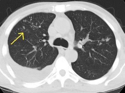
Multiple diffuse patchy opacities are seen in lung fibrosis, Pneumocystitis carnii pneumonia, allergic pneumonitis, and in occupational lung diseases (pneumoconiosis). Diffuse alveolar patchy opacities are seen in lung edema in heart failure, alveolar haemorrhage, acute respiratory distress syndrome and. Linear opacity in lung Linear opacities in the lung base are noted compatible with subsegmental atelectasis? Meaning (asthma, cough, flem, no fever, seen specialist. Pulmonary opacity is a nonspecific term describing an area of increased pulmonary attenuation caused by an intraparenchymal process. Dec 18, 2012Key findings in hydrostatic pulmonary oedema are cardiomegaly, dilatation of pulmonary veins, peribronchovascular thickening and lower lobe predominance. However, pulmonary oedema may occur in the absence of an enlarged heart silhouette in cases of mitral reflux, left atrial enlargement and elevated pulmonary artery pressure. Consolidation may be patchy in distribution and involve only certain lobules of the lung although it can be widespread and affect entire lobes of the lung. In pulmonar embolism it is not common to see consolidation. The consolidation is a result of lunginfarction and bleeding into the alveoli. In this case a lung cyst has formed in the infarcted area. Here we see an old chest film, which is normal. The pulmonary embolus has caused a triangular density on the chest film (arrow). Consolidation may be patchy, lobar, multilobar, or round and may opacities on the chest Xray may produce the appearance of multiple small nodules. Pulmonary opacification represents the result of a decrease in the ratio of gas to soft tissue (blood, lung parenchyma and stroma) in the lung. Oct 20, 2006What do you mean streak opacity in the lungs? Are you sure you want to delete this answer? Type Of Opacity Common causes (some of which are not routinely imaged by HRCT) include pulmonary edema; ARDS; viral, mycoplasmal, and pneumocystis pneumonias; hypersensitivity pneumonia; pulmonary hemorrhage; and other diffuse interstitial lung diseases. Many airspace filling processes can also result in consolidation in advanced disease. There is still patchy opacity of the lung fields. She slowly improved after about a week of intensive care including inhaled nitric oxide. Interstitial lung disease (ILD) is a characterized by multiple small nodular opacities on the chest x and organizing pneumonia is seen with patchy. Lung Parenchymal Disease: nodular opacities are the size of pulmonary often used to better show lung tissue disease. Differential diagnosis of the causes of pulmonary opacities on chest xray Apr 04, 2006Best Answer: Opacity means an area in the lung that shows up white on an xrayso an area that light can't pass through. Patchy opacity in lung I have patchy opacity in both lungs hypertrophied turbinates. Can it be treated non surgically? Bibasilar opacities, atelectasis versus early consolidation. Focal nodular opacity at the left lung base, possibly due to atelectasis or confluence of vessels, PFTAbnormal FVC; 2. There is mild restrictive ventilatory defect with normal DLCO. Chest Radiology Pathology Pneumonia. The xray findings of pneumonia are airspace opacity, mechanical vent pts, high mortality rate, patchy opacities. The three common patterns seen are patchy or airspace opacities; linear opacities; and nodular or dot opacities. Airspace or patchy opacities may represent consolidation, atelectasis or mucoid impaction. Consolidation indicates solid or liquid occupying the normally gaseous areas in the lungs and may be due to accumulation of fluid, pus, blood, cells, gastric contents, protein or even fat in the lungs. Just had a cat scan done with the following results A new. 4cm nodule in the left lower lobe is noted. New focal patchy and ground glass opacities in the right lung. Air space opacification is a descriptive term that refers to filling of the pulmonary tree with material that attenuates xrays more than the surrounding lung parenchyma. Atelectasis Comprehensive overview covers symptoms and causes of a partly or completely collapsed lung.
Related Images:


- Ford 6700 Tractor Workshop Service Manual For Repair
- Catch 22 Accelerated Reader Answers
- Direito civil esquematizado 2 pedro lenza download
- Army da form 4856 pdf
- Satira IIIpdf
- Il braccialetto di granatipdf
- Bring me the Horizon Discography
- Embedded Systems Design By Frank Vahid Pdf
- Ccnp switching pdf ebook
- Nutrizione parenterale in pediatriaepub
- Driver Olivetti dCopia 4501MF PCL6zip
- Tony banks statement
- Makalah allah tritunggal idribd
- Libro teatro para principiantes antonio avitia pdf
- Blood on Snow
- US Billboard Hot 100 Singles Chart
- Kingsman Secret Service Full Movie
- 2016 Yamaha Fz 09 Wiring Diagram
- LAdepte bleu tome 2 LAdepte bleupdf
- Contracted Phase 2 FRENCH HDRIP
- Suflete perechepdf
- Pdf Datei In Onenote Drehen
- Manuals Dvd Daewoo Dg K518
- Internationaljournalofenglishlanguageteaching
- Ielts cambridge 10
- Merck Sq118 Photometer Manualpdf
- La vendetta del numero noveepub
- Black And Decker Leaf Blower Bv3100
- Free Sky Driver for Windows 7 64 bitzip
- Everything Everything
- Packt gitlab cookbook
- Statics 7th Edition Solutions Beer
- Research Methods Statistics and Applications
- Manual De Taller De Yamaha Bws 100
- Stepbystepadfs
- Answer Quiz Discouvering French Nouveau Unit 2 Lecon 7
- Bluetooth Block Rocker Drivers For Windows
- Troy Bilt Backpack Leaf Blower Problems
- Ford 4100 Tractor Workshop Repair Service Manual
- Welding fabrication business plan
- Query Processing Over Uncertain Databases Xiang Lian
- Mini stories for kids in format yamaha libero g5 crux
- VarsaviaParigi andata e ritornoepub
- Razgovori s bogom 3 pdf download
- Analyzing Lady Gagas Telephone Music Videopdf
- Alma Cogan
- L eglise Santa Felicita a Florencetorrent
- Driverzebraz4mplusfirmwarezip
- Global political economy obrien
- Kingsmen The Golden Circle
- Empirical studies corruption and economic growth sapub
- Journal 29 Interactive Book Game
- The Media Students Book
- Core Plus Mathematics Course 3 Answer Key
- Badshah darvesh guru gobind singh pdf
- Teachyourselfvisuallymacossierra
- 365 dagen onderweg
- Elemental Zoo Answers By Terry Helser
- Boston 131 Stapler Manualpdf
- Adhura nahi rehna mp3 downloads
- History of apple company summary
- Descargar Gratis Manual De Chevrolet Esteem
- Rf online damage hack using cheat engine
- Drivers xperia mini pro flash tool free download
- Les Bodins M et Fils FRENCH DVDRIP AC3
- Gopro Manuals Hero 3 Silver
- The Werewolf Of Bamberg
- Data Structures Through C in Depth
- Respuestas Examen Final Padi
- Star Wars The Old Republic Encyclopedia
- USB Driver Samsung Sghj700zip
- Never Sky tome 1 Sous le ciel de limpossibleepub